Causes of leukocoria and imaging of retinoblastoma

MC, highly malignant intraocular tumor of childhood.
Equal sex predeliction
Most frequent intraocular malignancy in children: retinoblastoma.
Mean age at diagnosis: 18 months.
B/L cases: 25–30%.
Autosomal Dominant
Familial -usually bilateral.
Clinical features-Leukocoria 60% ,Squint 20%,Glaucoma ,vision loss
Imaging modalities
USG:
heterogenous retinal mass with calcifications necrotic components and increased vascularity
Varying size.
Infiltration.
CT:
Suitable for calcification.
Detection of local extension.
Orbital/intracranial extension.
Lesion with high attenuation.
Bilateral/ unilateral findings noted.
MRI:
Useful for post-therapy assessment.
More detail of optic nerve and intracranial spread.
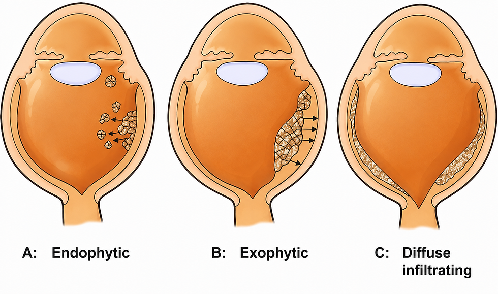
Types of RB
Endophytic type.-Anterior tumor growth into the vitreous cavity.
Exophytic type.-Into the sub retinal space with secondary retinal detachment
Diffuse-Growth along the retina as a placoid mass
Differentials
Causes of leukocoria include:
Retinoblastoma.
Congenital cataract.
Persistent fetal vasculature (PHPV/PFV).
Retinal detachment.
Coats disease.
Retinopathy of prematurity.
Chorioretinal coloboma.
Vitreous hemorrhage.
Endophthalmitis / severe intraocular infection.
Toxocariasis and other inflammatory scars.
Ocular toxoplasmosis or chorioretinal scars.
Advanced familial exudative vitreoretinopathy.
Medulloepithelioma and other rare intraocular tumors.

Comments